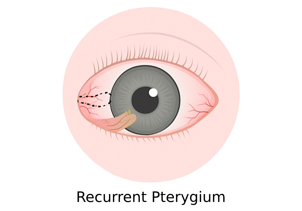
What is Pterygium?
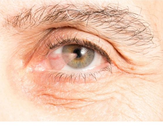
Pterygium is a non-cancerous, wing-shaped growth of tissue on the white part of the eye (sclera) that can extend onto the cornea. It usually forms on the side closest to the nose and may grow slowly over time.
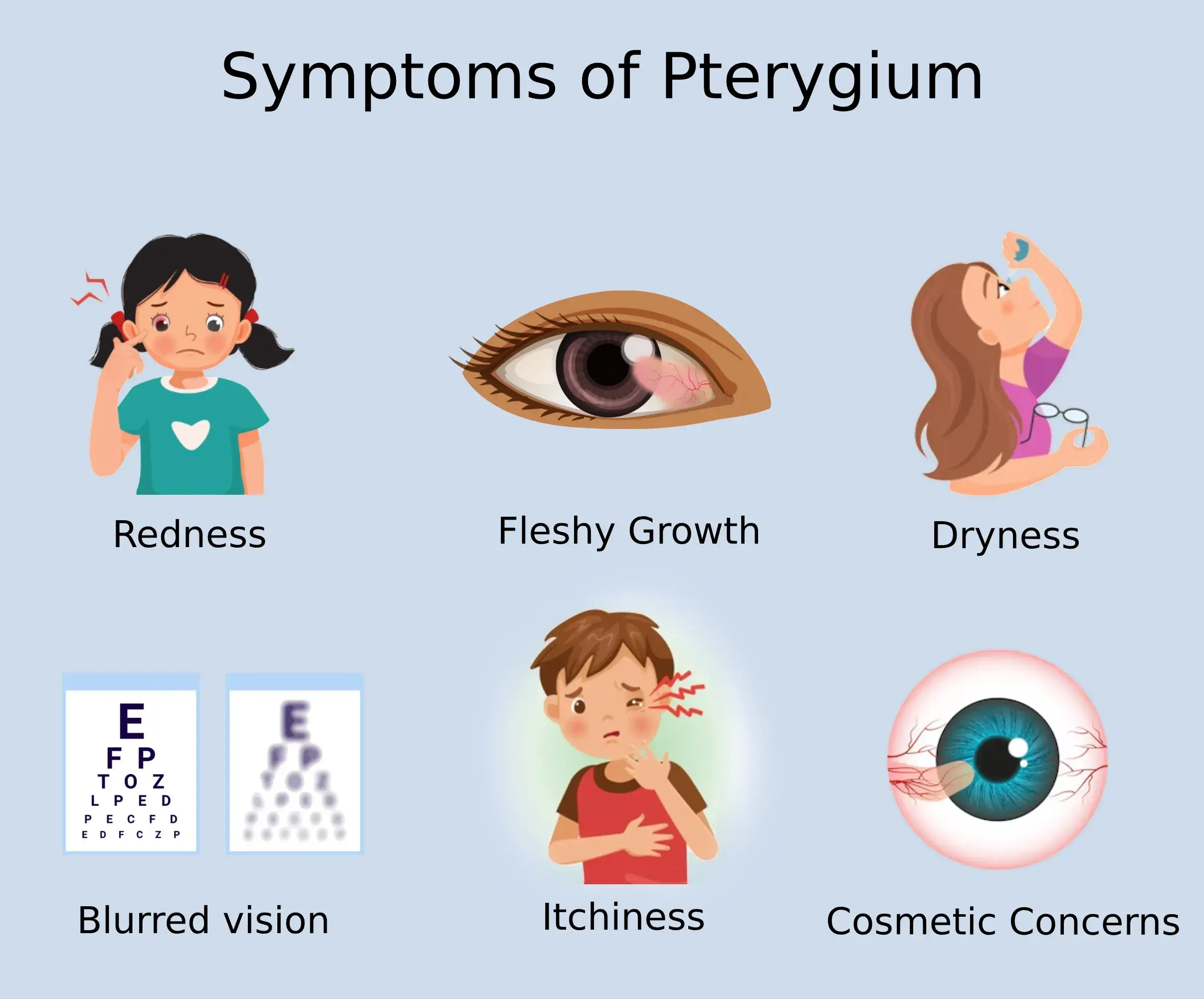
Common Symptoms of Pterygium
Pterygium may start without noticeable symptoms, but as it progresses, patients may experience various discomforts, including:
- Redness and eye irritation
- A visible fleshy growth on the white of the eye
- Foreign body sensation or gritty feeling
- Dryness and increased tearing
- Blurred vision (if the growth encroaches on the cornea)
- Itchiness or mild burning sensation
- Cosmetic concerns due to visible tissue on the eye
These symptoms may worsen with sun exposure, wind, smoke, or air pollution. Regular monitoring is essential to detect progression, especially if the pterygium begins affecting the cornea or altering vision. Artificial tears and protective eyewear can help manage mild symptoms before surgical intervention is needed.
.webp)
.webp)